

fig5
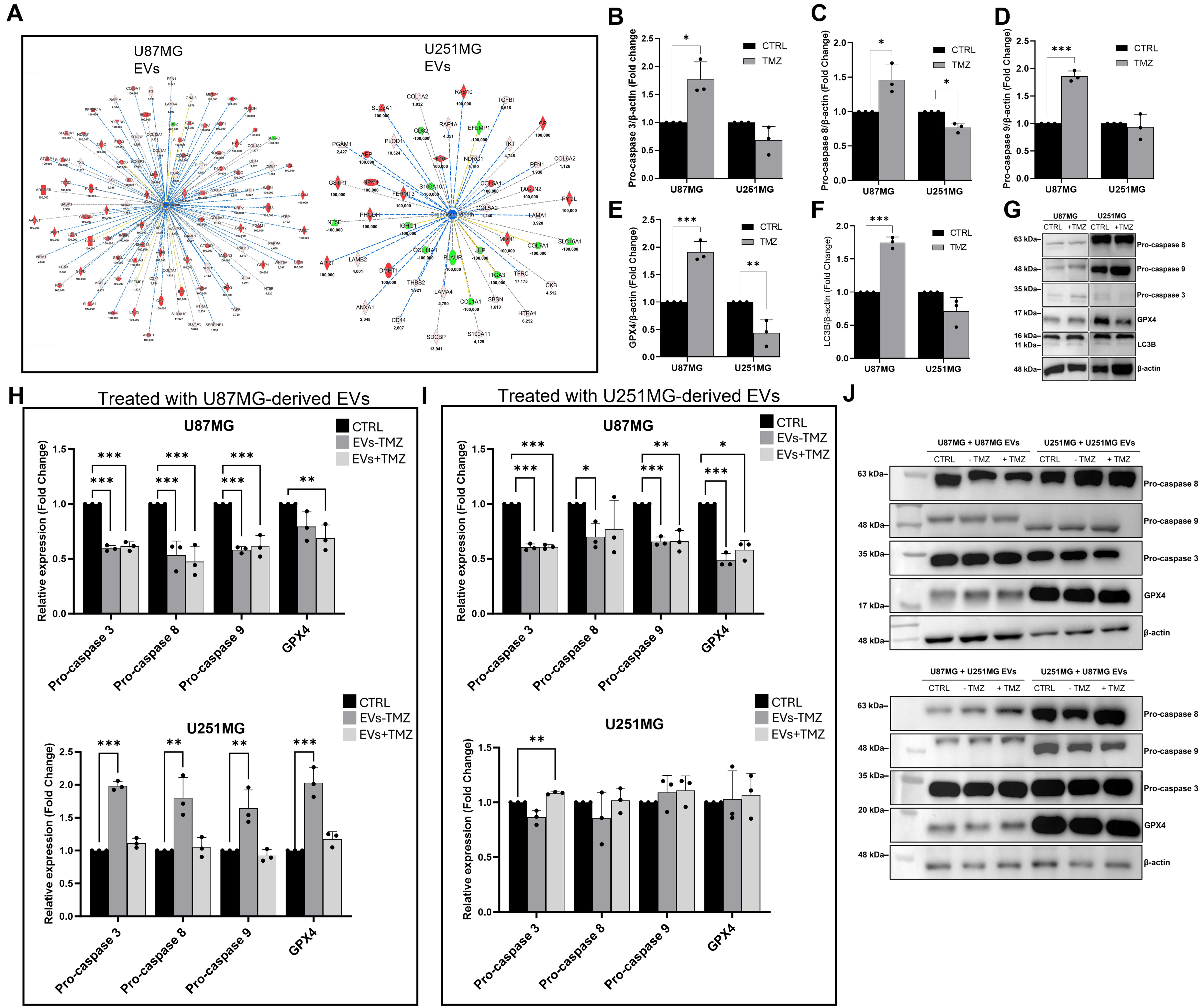
Figure 5. Effect of TMZ treatment on GBM-derived EVs and the expression of cell death- and stress-associated markers in TMZ-treated and EV-treated recipient GBM cells. (A) Downstream effect analysis of EV protein datasets revealed the inhibition of “Organismal Death” biofunction. Orange and blue shapes represent predicted activation or inhibition, respectively. Instead, red and green shapes represent increases or decreases in measurements of EV proteins, respectively, whose expression values are reported in the figure (ratio: EVs +TMZ/ EVs -TMZ). Continuous lines represent direct relationships, dotted ones represent indirect relationships, whereas grey and yellow lines indicate the non-predicted and inconsistent relationships. Expression levels of pro-caspase 3 (B), pro-caspase 8 (C), pro-caspase 9 (D), GPX4 (E), LC3B (F), and relative representative blots (G) of U87MG and U251MG cells untreated (control, CTRL) and TMZ-treated (TMZ) as determined by Western blot analysis; expression levels of pro-caspase 3, pro-caspase 8, pro-caspase 9, and GPX4 in untreated (CTRL) U87MG and U251MG and treated with EVs isolated from untreated (EVs -TMZ) and TMZ-treated (EVs +TMZ) U87MG (H) and U251MG (I) cells and relative representative blot acquisitions (J); Data are normalized to β-actin and expressed as fold change compared to the CTRL group. Statistically significant differences between groups were determined using one-way ANOVA followed by Tukey’s post hoc test ***P < 0.001. Data are presented as mean ± SD of three independent biological replicates (n = 3). Original and uncropped blots are reported in Supplementary Figure 5. ANOVA: Analysis of variance; CTRL: control; EVs: extracellular vesicles; GBM: glioblastoma; GPX4: glutathione peroxidase 4; LC3B: microtubule-associated protein 1 light chain beta; SD: standard deviation; TMZ: temozolomide.